Understanding Eye Anatomy: A Guide to How Your Eyes Work

For those who regularly use contact lenses, maintaining eye health is likely a top priority.
But have you ever wondered how vision actually works? It's often said that the eye functions like a camera, but what does that really mean?
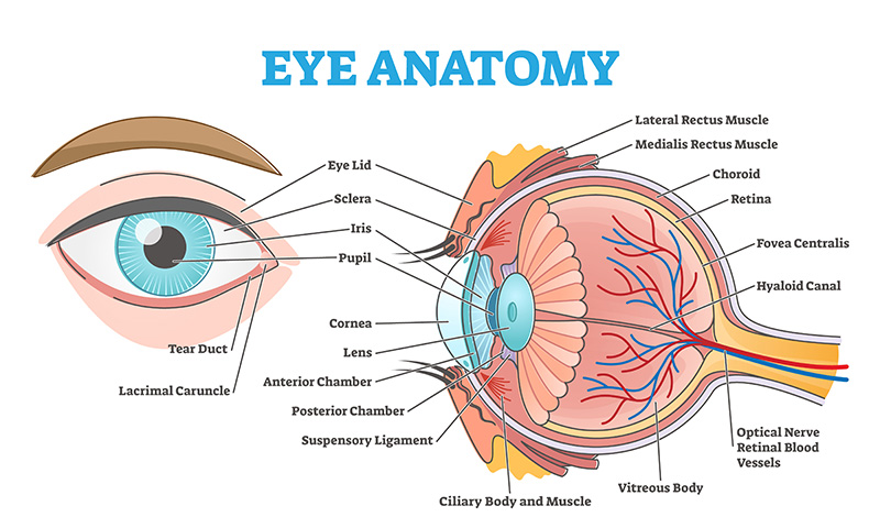
The eye is an organ responsible for vision, converting light into electrical signals and sending them to the brain. An adult's eye is a spherical structure about 24mm in diameter, made up of several parts. Light first passes through the cornea, then through the anterior chamber, the pupil, the lens, and the vitreous body before reaching the retina. The retina converts the light into electrical signals, which are transmitted to the brain via the optic nerve, allowing us to see.
It’s fascinating how the eye functions like a precision instrument, working seamlessly with the brain to create the images we perceive. In this article, we'll explore the anatomy and function of the eye and share tips on how to maintain optimal eye health.
Eye Anatomy and Function
Cornea
The cornea is the outermost, transparent, dome-shaped layer of the eye, covered by a thin layer of tears. It allows light to enter the eye and acts as a lens by refracting incoming light, accounting for about 70% of the eye's total refractive power. Additionally, the cornea helps protect the eye from germs, dust, and other contaminants. If the curvature of the cornea becomes irregular, it can lead to astigmatism, resulting in blurry vision.
Iris
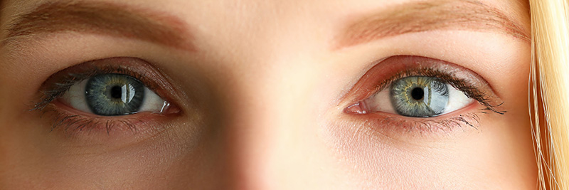
The iris is the coloured part of the eye visible through the cornea. It has a central opening called the pupil. The iris contains muscles that adjust the size of the pupil, similar to the aperture of a camera. By contracting or expanding these muscles, the iris regulates the amount of light entering the eye through the pupil. The colour of the iris can vary widely, with common categories including blue, green, grey, hazel, and brown.
Additionally, each person’s iris is as unique as a fingerprint, making iris scans highly effective for security purposes.
Lens
The lens is located just behind the iris and functions much like a camera lens within the eye, playing a crucial role in focusing light and adjusting the image.
Acting as a convex lens, it refracts light entering the eye and projects an image onto the retina, adjusting its focus as needed. When viewing distant objects, the lens becomes thinner, which reduces its refractive power. Conversely, when focusing on near objects, the lens becomes thicker, increasing its refractive power. This ability to change thickness allows the lens to focus on objects at varying distances.
As people age, they often develop presbyopia, a condition where the lens loses its ability to focus on close objects, making reading glasses necessary. Another common condition in older adults is cataracts, where the lens becomes clouded due to protein degeneration. The only effective treatment for cataracts is surgery, which is highly successful.
Retina
The retina is a thin, transparent membrane located at the back of the eye that detects light and forms images. After light passes through the cornea, iris, and lens, it is projected onto the retina. There, photoreceptor cells convert the light into electrical signals, which are then transmitted to the brain via the optic nerve. Due to this role, the retina is often compared to the film in a camera.
Optic Nerve
The optic nerve transmits visual information from the retina to the brain, where it is processed into images. This nerve has a complex structure, consisting of over a million nerve fibres. One common disease affecting the optic nerve is glaucoma. Glaucoma occurs when fluid accumulates in the eye, compressing the optic nerve and damaging nerve cells, usually due to increased intraocular pressure. As the pressure rises, the flow of aqueous humour is hindered, leading to compression and damage of the optic nerve. As the condition progresses, the field of vision narrows, and there is a risk of eventual blindness.
Occipital Lobe
The occipital lobe is the final stage in the visual process that begins when light enters the cornea of the eye. Images are converted into electrical impulses, which are then sent to the primary visual cortex in the occipital lobe. From there, the brain recognises objects and communicates with the temporal and parietal lobes to relate them to past memories and to perceive depth and three-dimensionality. Since sensory input from the eyes is continuous as long as they are open, the occipital lobe must operate at extremely high speed to keep up with the volume of information it receives. If the occipital lobe is damaged, a person may experience visual hallucinations or have difficulty recognising objects.
Other parts of the eye perform essential maintenance functions. While they do not directly assist in transmitting images to the optic nerve and occipital lobe, they are crucial for the overall functioning of the eyes.
Eyelids
The primary role of the eyelids is to protect the eyeballs through blinking. Blinking is essential for keeping the eyes moist and safeguarding them from foreign objects, external stimuli, and dryness. On average, we blink about 20 times per minute. However, when staring at a computer or smartphone for extended periods, the frequency of blinking decreases, along with tear production. This reduction in tears can lead to dryness of the eye surface, potentially causing "dry eye" and a decrease in vision. To maintain eye health, it’s important to consciously blink.

Eyelashes and Eyebrows
We often view eyebrows and eyelashes as decorative features, but they play crucial roles in maintaining eye health. Eyebrows help prevent perspiration from running down your forehead into your eyes and provide shade from bright light. Eyelashes protect the eyes by keeping foreign objects, such as dust, at bay. Additionally, the glands in the eyelashes produce sebum, which helps keep the inner surface of the eyelids lubricated.
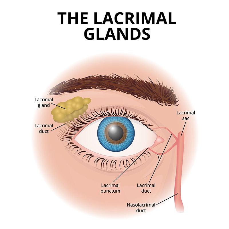
Lacrimal Gland
The lacrimal gland, located on the outer upper side of the eyelid, plays a crucial role in producing tears. Tears are secreted from the lacrimal gland and spread across the surface of the eye with each blink, helping to keep it moist and protect it from dryness. They are drained through small openings called lacrimal puncta at the inner corners of the eyelids and flow into the nose, while about 10% evaporates from the eye’s surface. Tears help maintain lubrication on the eye's surface, wash away foreign particles, and prevent infections. Proper functioning of the lacrimal gland is essential for eye health, as insufficient tear production can lead to dry eye syndrome.
Nasolacrimal Duct
The nasolacrimal duct is a tube that carries tears from the lacrimal sac to the nasal cavity. Tears discharged from the lacrimal puncta collect in the lacrimal sac and are ultimately drained into the nasal cavity through the nasolacrimal duct. Excess tears flow through the nasolacrimal duct into the inferior meatus of the nose. Under normal conditions (when you’re not crying), the tear ducts collect and drain these fluids into the nose. However, if the tear ducts are blocked, excess fluid remains in the eye, which can cause blurred vision and may lead to eye infections.
Even the most finely tuned machine doesn't always operate at its full potential and requires maintenance—this is especially true for our delicate eyes. Any issue with any part of the eye can severely impact vision and quality of life. That’s why it’s important to understand how the eyes work.
Today, there are many options available for treating vision loss caused by conditions of the cornea and lens. If you notice any abnormalities with your eyes, be sure to consult an ophthalmologist promptly and take appropriate measures to maintain your eye health. Additionally, even if you don’t have any eye issues, regular eye check-ups are essential.
For those who wear contact lenses, it’s crucial to use the correct lenses and take proper care of them to keep all parts of the eye healthy and functioning properly. By staying diligent with these practices and giving your eyes regular rest, you can maintain healthy eyes and clear vision for a long time.